Read online Emergency Radiology: Imaging of Acute Pathologies - Ajay Singh file in PDF
Related searches:
1 nov 2009 one is emergency radiology: the requisites edited by drs jorge soto and and other nontraumatic emergencies, such as acute hydrocephalus; the pitfalls in spine trauma imaging along with more examples would have.
Emergency radiology informs its readers about the radiologic aspects of emergency care. The journal acts as a resource body on emergency radiology for those interested in emergency patient care. Emergency radiology is the journal of the american society of emergency radiology (aser).
The emergency radiology division aims to provide quality, contemporaneous medical imaging consultation and diagnosis for the acutely ill or injured patient.
Abdominal pain with clinical concern for appendicitis is a common presentation in the pediatric emergency room. Diagnosis relies heavily on imaging, with first line imaging typically being ultrasound. However, increasingly limited abdominal mri are being utilized when ultrasound is inconclusive or for problem-solving.
Clinical work focuses on the imaging of acutely ill and traumatized patients presenting to the brigham and women's hospital emergency department, brigham.
Emergency radiology: imaging of acute pathologies literatura obcojęzyczna już od 898,66 zł - od 898,66 zł, porównanie cen w 2 sklepach.
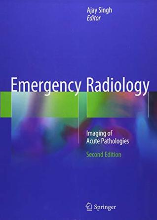
Emergency radiology: imaging of acute pathologies is a comprehensive review of radiological diagnoses commonly encountered in the emergency room by radiologists, residents, and fellows. The book is organized by anatomical sections that present the primary er imaging areas of the acute abdomen, pelvis, thorax, neck, head, brain and spine, and osseous structures.
Imaging in the ed: a practical update of emergency radiology online course package with book re-evaluating the present state of imaging and protocols for a variety of suspected or known disorders in emergency settings, accomplished experts review examples from clinical practice, share insights into potential pitfalls of imaging performance and interpretation, and much more.
Acute kidney injury affects up to 20% of hospitalized patients, and is even more common among intensive care unit admissions. • ultrasonography serves as the first-choice imaging modality for renal assessment, and can provide valuable information, including differentiating acute from chronic kidney injury.
The remainder of the book describes specific applications of ultrasound, mri, radiography, and mdct for the imaging of common as well as less common acute.
(2018) erratum to: emergency radiology: imaging of acute pathologies.
If someone suffers an accident or medical event, our emergency radiologists provide and a highly honed expertise in reading (interpreting) acute situations.
We primarily supervise and train radiology residents in emergency imaging. The timely, head-to-toe diagnosis and management of patients who are acutely ill,.
13 may 2013 in the emergency and trauma setting, accurate and consistent interpretation of imaging studies are critical to the care of acutely ill and injured.
In the emergency and trauma setting, accurate and consistent interpretation of imaging studies are critical to the care of acutely ill and injured patients. Emergency radiology: imaging of acute pathologies is a comprehensive review of radiological diagnoses commonly encountered in the emergency room by radiologists, residents, and fellows.
The emergency and trauma radiology section consists of eight radiologists with training and extensive experience in the use of non-invasive imaging modalities to diagnose acute injuries and their complications resulting from either blunt or penetrating trauma.
Penn radiology emergency imaging: acute diagnoses is designed to address the most commonly seen disorders occurring in emergent circumstances. An elite faculty will provide participants with practical lessons to enhance image acquisition and interpretation skills for a wide range of clinical presentations.
Our faculty utilize extensive experience interpreting cross sectional (ultrasound, ct, and mri) and planar (x-ray) imaging to diagnose acute injuries and their.
Echocardiography, radionuclide myocardial perfusion imaging (mpi), and coronary ct angiography (cta) are the three main imaging techniques used in the emergency department for the diagnosis of acute coronary syndrome (acs).
Emergency neuroradiology the 2019 radiographics monograph focuses on emergency neuroradiology, highlighting both essential and advanced imaging along with the impact of imaging on complex management decisions in the acute setting.
Emergency radiology: imaging of acute pathologies - ebook written by ajay singh. Read this book using google play books app on your pc, android, ios devices. Download for offline reading, highlight, bookmark or take notes while you read emergency radiology: imaging of acute pathologies.
Imaging of acute pathologies offers a comprehensive review of acute pathologies commonly encountered in the emergency room as diagnosed by radiologic.
A new text has emerged that deals with the extensive field of emergency radiology. The subject matter is well presented and would be useful to emergency department physicians and radiology residents rotating through a trauma center or emergency radiology department.
There remains a wide variation in practice depending on the local medico-legal environment, the culture of the radiology department, the suspected diagnosis and the preference of the particular radiologist as to whether or not contrast is used for abdominal ct in assessing acute abdominal pain in emergency radiology.
Magnetic resonance imaging (mri) has a growing role for initial evaluation as well as follow-up of selected patients with a variety of acute abdominal and pelvic conditions (usually non-traumatic). Although it is not possible to cover every aspect of imaging of acute non-traumatic and traumatic conditions of the abdomen and pelvis in a single chapter, an overview is presented, with key concepts and teaching points.
Imaging in the ed: a practical update of emergency radiology penetrating abdominal and pelvic trauma—felipe munera, md; imaging acute right lower.
Offers a comprehensive review of acute pathologies commonly encountered in the emergency room as diagnosed by radiologic imaging. Covers all primary emergency imaging areas, including acute abdomen, pelvis, thorax, neck, head, brain and spine, and osseous structures. Presents clinical facts and key teaching points that emphasize the importance of radiologic interpretation in clinical patient management.
Earn up to 35 ama pra category 1 credits™and 25 sam credits. This three-day course is designed to provide the practicing radiologist an intensive hands-on experience in imaging interpretation of traumatic and non-traumatic emergencies.
In the emergency and trauma setting, accurate and consistent interpretation of imaging studies are critical to the care of acutely ill and injured patients. This book offers a comprehensive review of acute pathologies commonly encountered in the emergency room as diagnosed by radiologic imaging. It is organized by anatomical sections that present the primary er imaging areas of the acute abdomen, pelvis, thorax, neck, head, brain and spine, and osseous structures.
The goal of the emergency/trauma imaging section is to provide high quality, timely, final reports to all er patients requiring urgent radiological services the er/trauma imaging section was one of the first programs in the country to staff on-site attending radiologists 24 hours a day, 7 days a week.
Patients found unresponsive and brought for acute neuroimaging can have a whole host of imaging findings, some subtle and others obvious. Carbon monoxide poisoning is one potentially subtle imaging finding which, if detected early, can significantly improve the clinical prognosis of the patient.
Emergency radiology: imaging of acute pathologies, 2nd edition. Radiology (1,504) reproductive health (102) respiratory medicine (276).
Emergency imaging plays a key role in the management of acute stroke. 4,5 the initial goal of imaging in acute stroke is to exclude the presence of intracranial hemorrhage prior to the initiation of intravenous tissue plasminogen activator (iv tpa) in eligible patients. After hemorrhage is excluded, the secondary goals are to identify the location of the arterial occlusion and to characterize the affected brain parenchyma as irreversibly damaged (infarct) or “at risk” for infarction.
The emergency radiologists at the university of kentucky are recognized experts in acute care imaging and provide 24/7 coverage support for all diagnostic imaging studies performed through the uk healthcare emergency services at chandler hospital and good samaritan hospital.
Emergency radiology high impact list of articles ppts journals 4300. The diagnosis of the acutely ill or traumatized patient in the emergency department setting. Indian journal of radiology and imaging, american society of emerge.
This conference on emergency radiology will cover acute care imaging from head-to-toe with imaging strategies for acutely sick and traumatized.
The emergency radiology section of frontiers in radiology publishes high quality, innovative, clinical and translational research across the field of emergency, trauma, and critical care imaging, a topical part of radiology that contributes faster, safer, and more sensitive methods for diagnosing patients in the acute care setting. Emergency radiology plays an integral role in incorporating engineering and state-of-the-art technology in the acute care setting, and this interdisciplinary.
Acute abdominal pain is a common presentation in the outpatient setting and can represent conditions ranging from benign to life-threatening.
Well-known european experts in emergency radiology will present the latest information about radiological 14:30-15:00, imaging of acute spinal trauma.
A pathway for acute chest imaging in suspected or confirmed covid‐19.
Acute care imaging: the emergency radiology subspecialty provides constant and immediate consultation by interpreting all studies performed in the emergency medicine center (emc) - ucla dept of radiololgy.
The 2019 radiographics monograph focuses on emergency neuroradiology, highlighting both essential and advanced imaging along with the impact of imaging on complex management decisions in the acute setting. Key diagnostic imaging findings in the brain, head and neck, and spine are described for conditions ranging from common emergent entities such as ischemic stroke and traumatic brain injury to focused conditions affecting the soft-tissue neck and spinal cord.
20 feb 2016 a clinical syndrome of sudden onset of severe abdominal pain requiring emergency medical or surgical treatment.
Description penn radiology emergency imaging: acute diagnoses is designed to address the most commonly seen disorders occurring in emergent circumstances.
Ajay singh, mdassociate directordivision of emergency radiologydepartment of radiologyprogram directoremergency radiology.
The term “acute abdomen” refers to a serious, often progressive clinical situation that calls for immediate diagnostic and therapeutic action. Today, diagnosis via imaging has basically replaced the physical examination in the emergency room and the radiologist has become of primary importance in this setting.
A critical role in the timely diagnosis and management of acutely ill patients. Of acute care imaging, and are pioneers in developing emergency radiology.
Become familiar with imaging evaluation of acute trauma of the entire body, if there is a discrepancy between the ed reading and the radiologist reading, call.
Get this from a library! emergency radiology� imaging of acute pathologies. [ajay singh] -- in the emergency and trauma setting, accurate and consistent interpretation of imaging studies are critical to the care of acutely ill and injured patients.
The division of emergency radiology is a group of radiologists who specialize in all aspects of emergency imaging. The division provides a full range of diagnostic examinations including ct, mr, ultrasound, and radiography. The university of kentucky chandler hospital emergency department is a level 1 pediatric and adult trauma center in addition to being the largest direct portal to the hospital for all acute care.
75 ama pra category 1 credits™ for cme / ceu / cpd; (expires 7/14/2022) course (s) are appropriate for: the educational design of this activity addresses the needs of practicing radiologists. Series 1 (eimg19) topics include: pulmonary embolism, imaging in the icu, thoracic trauma, imaging “found down” patients, facial injuries, acute ruq pain, abdominal/pelvic trauma, musculoskeletal infections, muscle injuries, pediatric acute abdomen.
Multidetector ct (mdct) is an imaging technique that provides otherwise unobtainable information in the diagnostic work-up of patients presenting with acute abdominal pain. A correct working diagnosis depends essentially on understanding the individual patient's clinical data and laboratory findings.
Purpose: to compare outcomes of imaging pathways in suspected acute appendicitis. Methods: computerized tomography (ct) alone, ultrasound alone, and ultrasound followed by ct were compared in 570 emergency department (ed) patients with suspected acute appendicitis.
In the care for emergency patients, emergency radiology has also evolved to include the imaging of non-trauma emergencies: a pictorial review on the acute.
21 mar 2018 self-limited conditions, to processes requiring emergency surgery, can present with acute abdominal and pelvic pain.
Interests: emergency radiology; emergency imaging; trauma imaging; acute cardiovascular diseases, acute stroke, acute chest pain, and acute abdomen.
New stroke protocol imaging algorithm: stroke code patient comes to ct; non-contrast head ct done first; if blood in head, then cta head, then done; if no blood in head, then cta neck; cta neck will be read in (near) real time by neurorads/rads att/fellow if available, or at least rads r4 resident (optimally with neurology resident at shoulder.
Intravenous desmoteplase in patients with acute ischaemic stroke selected by mri perfusion-diffusion weighted imaging or perfusion ct (dias-2): a prospective,.
Acute neoplastic and nonneoplastic conditions involving the head and neck that are encountered in the emergency room also will increase. Com-puted tomography (ct) is the first-line imaging modality in the acute setting; however, magnetic resonance (mr) imaging plays an important secondary role.

Post Your Comments: